The images tell a heartbreaking story: Zika’s calamitous attack on the brains of babies — as seen from the inside.
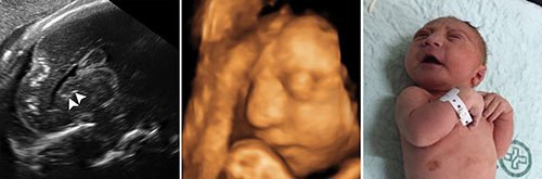
A study of brain scans and ultrasound pictures of 45 Brazilian babies whose mothers were infected with Zika in pregnancy shows that the virus can inflict serious damage to many different parts of the fetal brain beyond microcephaly, the condition of unusually small heads that has become the sinister signature of Zika.
The images, published Tuesday in the journal Radiology, also suggest a grim possibility: Because some of the damage was seen in brain areas that continue to develop after birth, it may be that babies born without obvious impairment will experience problems as they grow.

“It really brings to the forefront the importance of truly understanding the impact of Zika virus and the fact that we need to follow children who not only are exposed to Zika in pregnancy, but even those who don’t appear to have any complications at birth,” said Dr. Catherine Y. Spong, acting director of the Eunice Kennedy Shriver National Institute of Child Health and Human Development, who was not involved in the study.
Most of the babies in the study were born with microcephaly, although three were not. Each also suffered other impairments, almost all of which emerge earlier than microcephaly because a smaller head is really a consequence of a brain that has failed to develop fully or has been damaged along the way, experts said.
“The brain that should be there is not there,” said Dr. Deborah Levine, an author of the study and a professor of radiology at Harvard Medical School. “The abnormalities that we see in the brain suggest a very early disruption of the brain development process.”
The scans show the range of Zika’s brain targets, some of which experts knew about, including the corpus callosum, which facilitates communication between the two hemispheres; the cerebellum, which plays a significant role in movement, balance and speech; and the basal ganglia, which are involved in thinking and emotion.
“I think we were all aware that Zika causes brain abnormalities, but it’s been more generic,” said Dr. Rita Driggers, an associate professor of gynecology and obstetrics at Johns Hopkins University School of Medicine, who was not involved in the study. “Now we know more specifically what we’re looking for in terms of brain abnormalities before the microcephaly occurs.”
Continue reading the main story
Together, the images provide a more detailed guide that might help doctors diagnose Zika-related fetal damage earlier — possibly in the second trimester at a point early enough to help women decide whether to terminate a pregnancy, said Dr. Adre du Plessis, director of the Fetal Medicine Institute of Children’s National Health System, who was not involved in the study.
At the same time, the study may eventually help doctors rule out damage caused by Zika infection. “If there’s any uncertainty on ultrasound, we’re concerned that couples that are not risk-takers and don’t want to gamble might be terminating perfectly normal babies, which is of course a concern to us,” he said. “So there is a lot riding on being able to image accurately.”
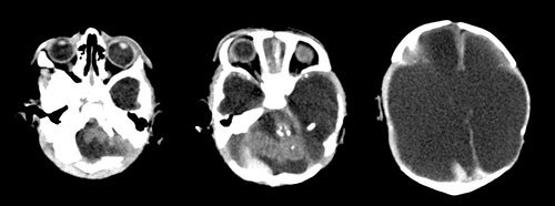
One finding that surprised several experts could become an especially meaningful diagnostic clue. Many infections that target the brain produce clumps of calcium, called calcification. But in Zika-infected babies, calcification often occurred in an unusual place: at the intersection of the gray matter of the outer layer of the brain, the cortex, and the white matter of the layer just below that.
That pattern could emerge as a particular stamp of Zika infection, experts said. Dr. Spong said that because the area involves two different types of blood vessels, it might suggest that Zika targets vascular areas.
And it could signal why the virus wreaks such ruthless effects on brain development.
“That is a critical area for brain formation,” Dr. du Plessis said. At the gray-white matter intersection, healthy cells “release certain chemicals that allow the neurons to find their precise destination.”
“When that gets scrambled they end up in the wrong place, they don’t function the way they should, and messaging and connectivity is severely deranged.”
Most of the babies in the study had such damage in the cortex, which plays a crucial role in learning, memory and coordination, and also continues to develop at least through infancy, suggesting that Zika-infected babies who seemed to emerge unscathed might be vulnerable to difficulties as they grow.
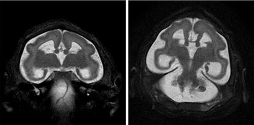
Another abnormality seen in most of the babies’ brains involved the ventricles or cavities of the brain becoming so full of cerebrospinal fluid that they “blow up like a balloon,” Dr. Levine said. The ventricles may be filling with fluid because Zika is obstructing their ability to drain normally, or because damage to other brain areas leaves a kind of vacuum that the enlarged ventricles fill.
The fluid-filled ventricles can make the head size seem normal earlier in pregnancy, Dr. Levine said. But as scans of one pregnancy taken at 36 weeks gestation show, the fluid can be so prominent that the scan shows what “looks like the skull and very little brain tissue inside it,” she said.
At some point, these ventricles, “like a balloon, can pop,” she said. And if they do, “the brain will collapse on itself.”
The images come from 17 babies whose mothers had confirmed Zika infection during pregnancy and 28 without laboratory confirmation but with all indications of Zika infection. Dr. Levine worked with colleagues in Brazil, which has more than 1,800 cases of microcephaly, to analyze images from the Instituto de Pesquisa in Paraiba in the northeastern part of the country. Three of the babies died in the first three days of life, and researchers studied autopsy reports in those cases.
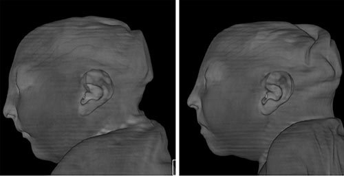
The images include scans of twin girls, who both developed microcephaly. The pictures show folds of overlapping skin and a sloping forehead, indications not only that the brain is smaller, but also that the forebrain has not developed normally, Dr. Levine said.
Images of another baby girl show contracted hands and arms, the result of another common symptom. Zika seems to damage the nerves in a developing fetus so that sometimes “muscles aren’t developing normally because they don’t have the nerve impulses to move normally,” she said. “And then when they’re born, they’re stuck in this contracted position.”
Dr. Levine said the images suggest that Zika is like a formidable enemy able to do damage in three ways: keeping parts of the brain from forming normally, obstructing areas of the brain, and destroying parts of the brain after they form.
With such a vicious and unpredictable virus, “it’s key to realize that Zika is more than microcephaly, that there’s a number of other abnormalities as they’ve shown in this paper, and its effects are going to be even more broad,” said Dr. Spong, whose agency has begun a study of what will ultimately be 10,000 babies born in Zika epidemic areas including Brazil and Puerto Rico.
“It’s going to be essential to follow them to look at their development, to look at their ability to learn, to look at hearing problems, balance problems, behavior problems, all those issues, to make sure that we don’t miss anyone.”
Source: www.nytimes.com